Introduction
Functional foods contain bioactive compounds that provide physiological and health benefits beyond nutrition, and the emergence of these foods has led people around the world to gradually transition to a diet rich in proteins, essential oils, vitamins, polyphenols, and carotenoids (3 Angeles et al., 2021). Among the major bioactive constituents of functional foods, proteins and peptides have attracted considerable attention because of their diverse physiological activities. Proteins play a fundamental role in providing the nutritients for human diet derived from foods. Proteins derived from functional foods may exhibit various health benefits, including regulation of blood pressure, cholesterol levels, and blood sugar.
This growing interest in functional foods is largely associated with the increasing prevalence of chronic non-communicable diseases, particularly cardiovascular diseases (CVD). Two major modifiable risk factors of CVD are hypertension and dyslipidemia. Hypertension affected around 1.3 billion adults in 2023, while dyslipidemia accounts for approximately 4 million CVD-related deaths worldwide (Zhao, 2021; World Health Organization, 2023; Abera, Worede, Hirigo, Alemayehu, and Ambachew, 2024). Increased consumption of plant proteins has been associated with reduced CVD risk. Due to this emerging need of the populace, the development of nutritionally enhanced and functional foods to address health concerns is rapidly increasing.
The physiological effects exhibited by proteins are usually present in the peptide sequences encrypted in the parent protein, which become active when released as peptide fragments or free amino acids. Bioactive peptides found in foods have been widely investigated for their potential role in the prevention of conditions such as hypertension, cancer, dyslipidemia, microbial infections, and cardiovascular diseases (Cicero, Floor and Albertoni, 2017). Protein hydrolysates and peptides from cereals, legumes, and animal products have been widely investigated for their ACE-inhibitory and lipid-lowering activities, yet fruits are generally overlooked as sources of proteinaceous compounds due to their relatively low protein content. This gap underscores the need to explore unconventional fruit sources using targeted workflows that combine protein extraction, characterization, digestion, and in vitro evaluation aligned with antihypertensive and lipid-related outcomes.
Calabash (Crescentia cujete L.) is of particular interest because its fruit has long been used in traditional medicine and has recently gained considerable attraction in the Philippines due to anecdotal claims of treating hypertension, diabetes, dyslipidemia and cancer. Belonging to the family Bignoniaceae, calabash is a seasonal fruit that is widely propagated usually in home gardens in Central and South America, Africa and Asia. Calabash fruit has been incorporated into herbal mixtures in many traditional medicine systems for its anti-inflammatory, and anti-bacterial effects and its use in managing cough and colds (Shastry, Bhalodia Maulik and Aswathanarayana, 2012). Although the biological activities of calabash are commonly attributed to phytochemicals such as apigenin, quercetin, anthraquinones, carotenoids, and β-sitosterol, the contribution of its proteinaceous components remains poorly understood.
In this context, the present study aims to isolate, purify and characterize the proteins and peptides released after simulated gastrointestinal digestion of calabash fruit flesh, thereby providing baseline information on its protein content. This study also aims to investigate the potential effects of proteins and peptides in managing elevated blood pressure and high cholesterol levels, providing scientific validation for anecdotal claims.
Materials and Methods
Sample Preparation and Crude Protein Isolation
Calabash (C. cujete) fruits were collected from the province of Davao, Philippines. Three randomly selected trees were used as biological sources, and three mature fruits were harvested from each tree yielding a total of nine fruits for subsequent analysis. Fruit maturity was determined based on fruit size, hard outer rind and external color, which are used for morphological identification of mature calabash fruits. The fruit flesh was lyophilized using a -85˚C laboratory floor-mode freeze dryer (IlShin Biobase, Korea). Lyophilized fruit flesh from each fruit was extracted individually for generate nine independent samples, and downstream assays were performed in triplicate for each extract. Two grams of lyophilized calabash flesh were homogenized with 10 mL of 0.1 M sodium phosphate buffer pH 7.0 containing 0.05 M Tris-HCl and 0.4 M NaCl. The mixture was incubated overnight at 4˚C with shaking, and then filtered through cheesecloth and centrifuged at 11,872 x g at 4˚C for 10 min. The resulting filtrate was labeled as crude protein isolate (CPI).
Purification of Proteins
Ammonium Sulfate Precipitation
Five mL of CPI was subjected to ammonium sulfate precipitation to achieve approximately 40% saturation by slow addition of 1.21 grams of powdered ammonium sulfate under constant stirring. This saturation level was selected, based on preliminary experiments, to recover a protein-enriched fraction. The samples were centrifuged at 11,872 x g for 10 min at 4˚C using Beckman Coulter X-30R (USA). The supernatant was discarded, and the pellet was reconstituted using 0.1 M sodium phosphate buffer pH 7.0.
Dialysis
The reconstituted proteins were dialyzed overnight at 4˚C against distilled water using a cellulose dialysis tubing with molecular weight cut-off of 3.5 kDa. The distilled water was changed every 3 h.
Gel Filtration Chromatography
Dialyzed protein samples were purified using Sephadex G-100 (Sigma-Aldrich, USA) pre-swollen for 3 days and equilibrated using extraction buffer. Three milliliters of dialyzed protein samples were loaded into the column and eluted with cold extraction buffer at a flow rate of 1.0 ml/min. Fractions of 1 mL each were collected, and absorbance of eluates was monitored at 210 nm using a UV-Vis spectrophotometer (Spectroquant Pharo 300, Merck Millipore, Germany). Pooled fractions were labelled as purified proteins (PP).
Protein Quantification Using Bradford Assay
Protein quantification was performed using Quick Start Bradford Assay (Bio-Rad, USA). A standard calibration curve was prepared using different concentrations of bovine serum albumin (BSA). In a 96-well microtiter plate, 2.5 µL of sample and 125 µL 1x Bradford reagent were thoroughly mixed and incubated for 10 min. The absorbance was monitored at 595 nm using a microplate reader (BioTek Epoch, Agilent, USA).
Sodium Dodecyl Sulfate–polyacrylamide Gel Electrophoresis (SDS-PAGE)
Twelve percent (12%) discontinuous denaturing gels were prepared and run at 110V for approximately 100 minutes using a Mini-PROTEAN Tetra Cell (Bio-Rad, USA). Gel bands were visualized using 0.1% Coomassie Brilliant Blue R-250 (Bio-Rad, USA). A protein reference ladder (NEB, MA, USA) was used to estimate the molecular weights of the protein subunits.
Enzymatic Hydrolysis of Proteins
PP were subjected to digestion in a 12:1 protein-to-enzyme ratio. Solutions of pepsin, trypsin and chymotrypsin (1 mg/mL) were prepared in distilled water. These enzymes were chosen to simulate the main stages of human gastrointestinal protein digestion and to generate a broad spectrum of peptide fragments with different sequence specificities under physiologically relevant pH and temperature conditions. Sequential digestion was performed, wherein pepsin was added first and allowed to digest for 1 h at pH 2.0. The remaining enzymes were then added simultaneously after adjusting the pH to 7.0 using 1.0 M NaOH. The mixture was incubated for 1, 2, 3, 4, 6 and 24 h at room temperature. The hydrolysis was terminated by immersing the samples in a boiling water bath for 5 min. Progression of hydrolysis was assessed via SDS-PAGE.
Normalization of Protein Activity
The normalized protein activity of CPI was obtained by dividing the percent inhibition by the mass of protein loaded per assay. For CPI, the protein mass was calculated from the Bradford-derived concentration (688 µg/mL) and the assay loading volume, and the values are expressed as apparent normalized inhibition per mg protein. Activities were therefore expressed as apparent normalized percent inhibition per mg protein loaded. Because the purified fractions fell below the Bradford detection limit, normalization of PP digest activity should be considered approximate unless verified using a more sensitive protein assay.
In vitro Assays for Antihypertensive, Anti-obesity and Hypocholesterolemic Activities
The sample preparation and laboratory analysis were all done at the Biochemistry Laboratory of the Institute of Plant Breeding, College of Agriculture and Food Science, University of the Philippines Los Baños. All chemicals and reagents used were of analytical grade and purchased from reputable suppliers.
ACE Inhibition Assay
ACE inhibition activity was determined using the method of Cushman and Cheung (1971) with modifications. An assay mix of 25 μL 100 mM sodium phosphate buffer pH 8.3, 25 μL 300 mM NaCl, 50 μL 5 mM hippuryl—histidyl-leucine (HHL), 25 μL of sample, and 25 μL crude ACE extract (which was added last) from pig lungs was incubated for 30 min at 37˚C. After incubation, 125 μL of 1 N HCl was added to stop the reaction. A blank control was prepared by adding 25 μL of phosphate buffer to the assay mix instead of the sample. Captopril at a concentration of 1 mg/mL was used as a positive control and added to the assay mix. Negative control was also prepared wherein the stop solution (1 N HCl) was initially added to the assay mix without ACE. Addition of 750 μL ethyl acetate into the mix was then performed to extract hippuric acid. Five hundred microliters of the ethyl acetate layer were collected and allowed to evaporate to dryness. The extracted hippuric acid was dissolved in 500 μL distilled water and the response was monitored at absorbance of 228 nm. The percent inhibition was calculated using the following equation:
Pancreatic Lipase Inhibition Assay
The pancreatic lipase inhibition activity was determined using the method of Chheda, Kaikini, Bagle, and Seervi, (2016). Freshly prepared 0.1 M potassium phosphate buffer pH 7.2 with 150 mM NaCl and 0.5% (v/v) Triton X-100 was used to dissolve 6 mg of lipase enzyme from porcine pancreas (≥125 units/mg protein) and diluted to 10 mL with phosphate buffer. The substrate of the assay was prepared using 8.45 µL p-nitrophenyl butyrate (PNPB) and diluted to 10 mL with acetonitrile. Inhibitory activity was determined by mixing 25 µL of the sample, 100 µL phosphate buffer, 50 µL lipase enzyme, and 25 µL PNPB solution. The mixture was incubated at 37˚C for 30 min and the absorbance was monitored at 400 nm using a microplate reader to estimate the amount of p-nitrophenol released from the reaction. One mg/mL Orlistat dissolved in dimethyl sulfoxide was used as a positive control. The percent inhibition was calculated using the following equation:
Cholesterol Micellar Solubility Inhibition Assay
The cholesterol micellar solubility inhibition activity was determined using the method of Bangoura, Wenshui, and Jiali, (2009). Cholesterol micellar solution was initially prepared by mixing 10 mM sodium deoxycholate, 0.4 mM cholesterol, 132 mM NaCl, 1 mM oleic acid, , and 15 mM potassium phosphate buffer (pH 7.4). The mixture was sonicated before use.
A 1:1 mixture of micellar solution and sample was incubated at 37˚C for 24 h and centrifuged at 11,872 x g for 10 min. Using a 96-well titer plate, 60 µL of supernatant was mixed with 75 µL of acetic acid followed by the slow addition of 90 µL Zak’s reagent (0.1% FeCl3 in concentrated H2SO4). The reaction mixture was incubated at room temperature for 30 min prior to absorbance determination at 560 nm using a microplate reader. One mg/mL cholestyramine dissolved in phosphate buffer was used as positive control. The blank sample was prepared by substituting the protein isolates with the micellar solution. The percent inhibition was calculated using the following equation:
Statistical Analysis
The data collected were expressed in mean ± standard deviation and analyzed using R Studio (Posit Software, Boston, Massachusetts). The number of samples, n, refers to independently prepared fruit extracts, whereas assay measurements from each extract were treated as technical replicates. The bioactivities were analyzed using Kruskal–Wallis test to determine the significant differences among the given groups. A pairwise post hoc test using Dunn’s test with Benjamini-Hochberg (BH) correction was also performed to identify which specific groups differ compared to the activity of positive control.
Results and Discussion
Protein Isolation and Characterization
This study extracted soluble protein-containing fractions from C. cujete fruit flesh to evaluate their functional applications and potential health benefits through in vitro bioactivity assays. The extracted protein has an average concentration of 688 µg/mL in terms of extraction buffer or 0.344 g/100 g dry weight of fruit flesh. The median and interquartile range (IQR) values were recorded at 671 and 449 µg/mL, respectively. The quality of CPI was assessed using SDS-PAGE as shown in Fig. 1a. Five distinct bands were observed with estimated molecular weights of 12.1 kDa, 23.2 kDa, 25.7 kDa, 30.1 kDa, and 46.7 kDa. This suggests that CPI contains relatively low-molecular-weight proteins and peptides. Many classical structural and enzymatic proteins in cellular compartments have higher molecular masses (usually 40–60 kDa), but numerous functional proteins and bioactive peptides are substantially smaller. Classic examples include insulin, a 5.8 kDa hormone, and the 4-kDa hormone-like legume peptide that interacts with a 43-kDa receptor-like kinase in soybean. Plant-derived proteolytic enzymes like fruit bromelain (25-32 kDa) from pineapple, widely used for its protease activity and reported anti-inflammatory property further demonstrate that functional proteins can span a broad molecular-weight range. Reviews on plant- and food-derived bioactive peptides also report many active sequences with molecular weights below 10 kDa, supporting the view that the relatively small size of the CPI proteins and peptides observed in this study is compatible with functional and bioactive roles (Hanada and Hirano, 2004; Hikisz and Bernasinska-Slomczewska, 2021; Mayer, Zhang, and DiMarchi, 2007; Zhuet al., 2023).
Crude proteins were isolated using sodium phosphate buffer containing Tris-HCl and NaCl. Phosphate buffers are highly effective in maintaining pH stability, thereby preventing extreme changes that could result in protein denaturation or aggregation. Maintaining a neutral pH (7.0) is crucial for preserving the native conformation of proteins, ensuring their functionality and bioactivity remain intact. Furthermore, sodium phosphate buffer is a preferred choice in protein extraction protocols due to its minimal reactivity with proteins, which reduces the likelihood of undesirable chemical interactions (Pavani, Kumar, Rani, Venkatesu, and Lee, 2021). CPI contains less than 1% protein, consistent with the typically low protein content of most fruits and vegetables, which still offer dietary benefits (Fordet al., 2023).
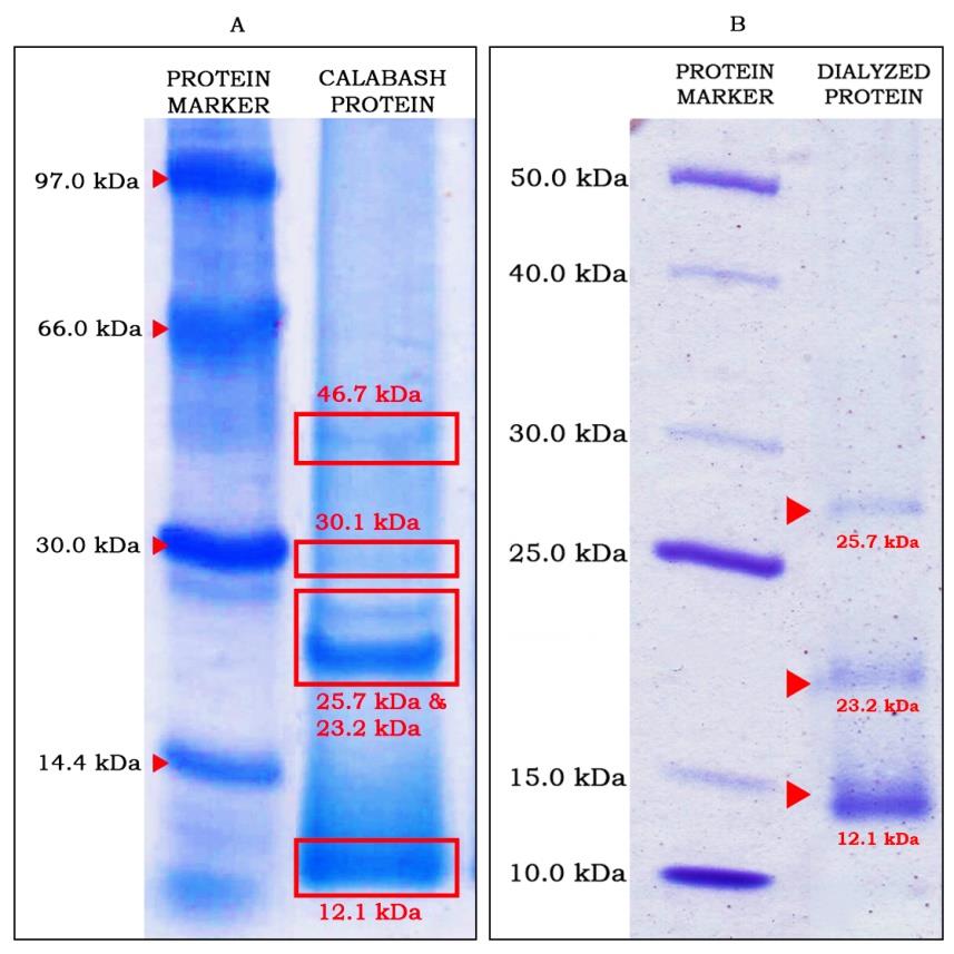
Fig. 1. Electrophoretograms of (a) CPI highlighting five major protein bands; and (b) partially purified proteins from ammonium sulfate fractions
The CPI was precipitated using ammonium sulfate at 40% saturation as a partial enrichment step and subsequently dialyzed to remove excess salts. This precipitation indicates selective recovery of a subset of soluble proteins rather than confirmed precipitation of the total protein complement. Classical protein fractionation shows that proteins precipitate over a broad range of saturations (e.g., 60-80%) to recover more complete protein fractions (Tola and Missihoun, 2023; Wingfield, 1998). Future work will combine stepwise fractionation with trichloroacetic acid or total nitrogen measurements to validate total protein recovery. The partially purified proteins were evaluated using SDS-PAGE which showed three remaining distinct bands with improved resolution (Fig. 1b) which correspond to molecular weights of 12.1 kDa, 23.2 kDa, and 25.7 kDa, as estimated in CPI in Fig. 1a. Different molecular weight protein markers were used for Fig. 1a and 1b due to reagent availability during separate experimental runs; however, band size estimations were independently performed using the corresponding protein ladder for each gel. The partially purified proteins were then subjected to size-exclusion chromatography using Sephadex resin and 25 fractions of 1 mL each were collected. The elution profile showed two distinct peaks: one between 5-9 mL and another broad peak from 11-22 mL with observed peak shouldering from 15 mL to 18 mL. The presence of shouldering suggests the presence of multiple species due to close molecular weight or the existence of multiple states such as oligomer or protein aggregates. After conducting several electrophoretic runs, the remaining visible band corresponded to a protein of approximately 12.1 kDa, matching the last broad peak observed in the elution profile as illustrated in Fig. 2. Bradford assay could not determine the remaining protein concentration in PP because of the possibility of having few low molecular weight proteins and peptides. Another possibility is that the protein concentration of PP, estimated to be less than 20 µg/mL, is already below the detection limit of the Bradford assay, thus resulting in no detectable response. Other protein quantification methods with higher sensitivity and detection limit such as Lowry and Bicinchoninic Acid (BCA) assays can be utilized in the future to verify the protein content of calabash fruit flesh.
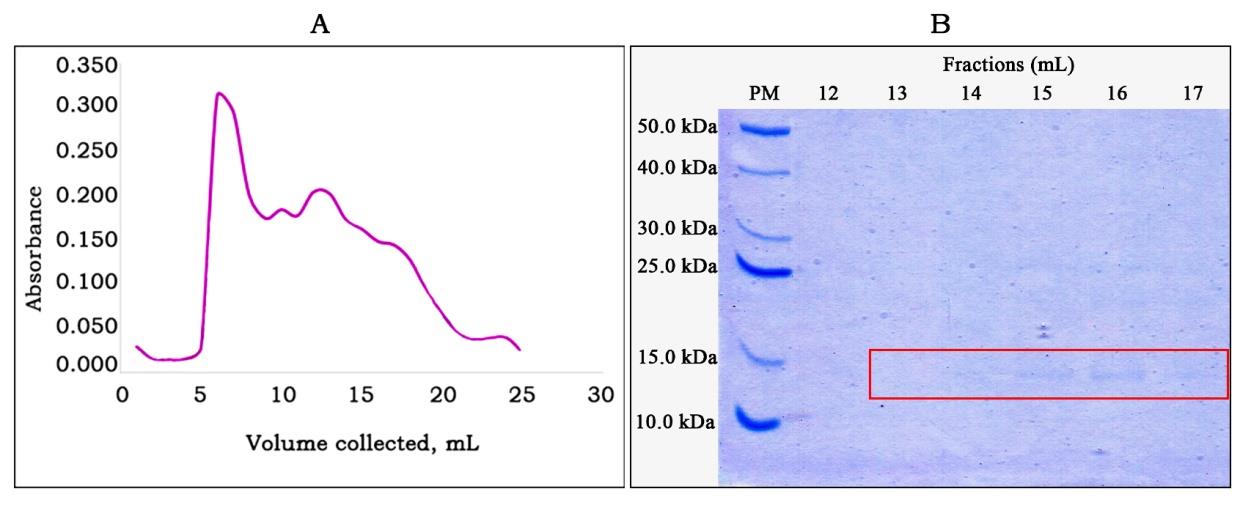
Fig. 2. (a) Elution profile of the dialyzed 40% ammonium sulfate-recovered protein fractions, and (b) SDS-PAGE profile of PP with visible bands with approximate molecular weight of 12.1 kDa
PP was used to simulate gastrointestinal digestion. Enzymatic digestion releases peptides and amino acids that were initially inactive within the native protein structure. These peptides are known to exhibit protein-protein inhibition due to their variability in size and specificity (Daliri, Oh and Lee, 2017). After 1 h of digestion, protein bands were no longer visible in the polyacrylamide gels, indicating successful protein hydrolysis resulting in low molecular weight proteins or peptide fragments. This observation suggests the efficiency of the hydrolysis process and highlights its potential to support biochemical reactions analogous to natural digestive processes. In addition, purified protein hydrolysates were not detectable by Bradford assay, likely due to low concentration or reduced dye binding of low-molecular-weight species, hence, the extent of total protein recovery could not be fully verified in the present study.
The apparent normalized inhibition values reported for CPI (Table 1) represent protein-normalized endpoint responses rather than classical enzyme specific activities, which are typically expressed in units per mg based on kinetic measurements. Because the purified protein fraction and its hydrolysates could not be reliably quantified by Bradford, equivalent normalization for these samples was not performed, and their activities are presented as percent inhibition per reaction as presented in Table 2.
| Apparent % inhibition per milligram protein | |||
|---|---|---|---|
| ACE inhibition | Lipase inhibition | Cholesterol micellar inhibition | |
| Crude Protein Isolate (CPI) | 4.45 × 10³ | 1.30 × 10³ % | 2.03 × 10³ % |
| ACE inhibition, % | Lipase inhibition, % | Cholesterol micellar inhibition, % | ||||
|---|---|---|---|---|---|---|
| CPI | PP digest | CPI | PP digest | CPI | PP digest | |
| Positive control | Captopril 78.9 ± 5.94a | Orlistat 72.6 ± 6.36a | Cholestyramine 91.5 ± 0.16a | |||
| Calabash | 76.5 ± 3.54a,b | 73.4 ± 1.00a.b | 22.3 ± 5.76b,c | 44.9 ± 0.680a,c | 84.0 ± 1.20a,b | 82.2 ± 8.17a,b |
| *Values are expressed as mean ± SD (n = 3). In the same column, values followed by the same superscript letters are not significantly different at the 5% level of significance based on the Kruskal-Wallis test followed by Dunn’s test. Letters before the comma indicate column-wise comparisons; letters after the comma indicate row-wise comparisons per bioassay | ||||||
ACE Inhibition Activity
The in vitro antihypertensive and lipid-lowering properties of CPI and PP hydrolysates were determined spectrophotometrically. For antihypertensive activity, CPI exhibited high ACE inhibition of 76.5%, which showed no significant difference from Captopril, a known ACE inhibitor drug, with 78.9% inhibition (p>0.05). CPI contributed approximately 0.0172 mg protein per reaction, corresponding to an apparent normalized inhibition of about 4.45 × 10³ % inhibition per mg protein. This value provides a protein-normalized estimate of the ACE inhibitory response of CPI and supports the observation that the crude isolates contain proteinaceous components with inhibitory potential. However, this apparent normalized inhibition should be interpreted only as an internal comparative metric because the assay was performed as an endpoint inhibition assay rather than a kinetic assay. In addition, inhibition values of the calabash CPI approached those of captopril under the assay conditions, such comparisons should be interpreted cautiously because the tested materials differed markedly in composition, purity, and active mass. In addition, results showed that ACE inhibition of protein hydrolysates had comparable values from the 1st to 4th h of digestion but showed a significant decrease at the 6th h of digestion. However, an increase in inhibition activity was later observed for the 24-h digests. For visualization, the effect of digestion on the purified samples over time using ACE inhibitory activity is shown in Fig. 3.
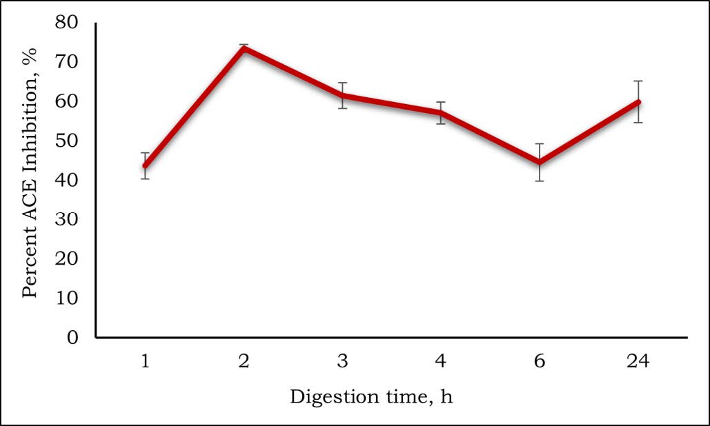
Fig. 3. Effect of digestion time on ACE inhibition of C. cujete fruit flesh purified protein hydrolysates
For digested PP, it was observed to have high ACE inhibition after 2 h digestion time with 73.4% inhibition compared to other digestion times. The 2-h digests were selected to use in the succeeding bioassays after the ACE inhibition assay exhibited the highest bioactivity across digestion time.
Crude extracts may contain various proteins in their native conformation including the presence of other bioactive compounds such as polyphenols, which could contribute to the observed increased bioactivity. Previous studies have shown that certain phenolics and flavonoids exhibit ACE inhibitory activity (Balasuriya and Rupasinghe, 2011; Simaratanamongkol, Umehara, Noguchi, and Panichayupakaranant, 2014). The results obtained from this study were comparable to the ACE inhibitory activity of mango flesh and ‘Señorita’ banana cultivar flesh crude proteins with 71%-74% and 85.2% inhibition, respectively (Ferreras, Bercansil-Clemencia, Hizon-Fradejas, Uy, and Torio, 2021; Rojaset al., 2021). The highest ACE inhibition was observed in 2 h digested PP, followed by a significant decrease at the 6th h of digestion. Although the 2-hour digest exhibited highest ACE inhibition, the present study did not fractionate the hydrolysate into peptide subfractions. The initial increase in activity likely reflects the release of ACE-inhibitory peptides during early digestion time whereas the subsequent decrease may be attributed to the further hydrolysis of proteins into smaller fragments with reduced inhibitory response. ACE inhibitory peptides are typically short sequences (2–12 amino acids), because smaller peptides can more easily access the ACE active site. Their activity is commonly associated with hydrophobic or aromatic residues, especially at the C-terminal end. These features are inferred to enhance binding through hydrophobic interactions within ACE’s substrate-binding pockets leading to an improved inhibitory response (Daskaya-Dikmen, Yucetepe, Karbancioglu-Guler, Daskaya, and Ozcelik, 2017; Huet al., 2025). ACE inhibition activity can also be increased by using higher enzyme concentration and longer hydrolysis time which may generate shorter peptides with potential bioactivity (Rojaset al., 2021; Shalaby, Zakora, and Otte, 2006). Similar studies also reported higher ACE inhibitory activity when exposed to longer hydrolysis time (Abuine, Rathnayake and Byun, 2019). In addition, a minimal contribution can be linked to trace amounts of other compounds, such as polyphenols because PP underwent purification and dialysis utilizing a molecular weight cut-off of 3 kDa which effectively removes compounds smaller than this threshold. Any residual effect would therefore be minor compared to peptide-mediated inhibition.
Pancreatic Lipase Inhibition Activity
For pancreatic lipase inhibition, CPI exhibited 22.3% inhibition and showed significantly lower inhibition than Orlistat, a known lipase inhibitor, with an inhibition value of 72.6 %. The crude protein isolate corresponded to an apparent normalized inhibition of approximately 1.30 × 10³ % inhibition per mg protein. This lower normalized response relative to ACE and cholesterol micellar solubility inhibition suggests that, within CPI, lipase-inhibitory activity was weaker or less concentrated than the other measured bioactivities. On the other hand, 2-h PP digest showed higher lipase inhibition at 44.9% compared to CPI and was not statistically significant from Orlistat (p>0.05). The values obtained in this study were lower compared to Orlistat but higher compared to the reported lipase inhibitory activity of sea buckthorn seed protein hydrolysates with 35% inhibition (Xianget al., 2020). Studies also reported peptides isolated from millet grains, rice bran, milk, and some fish species that showed moderate to strong inhibitory activity (Jakubczyk, Szymanowska, Karaś, and Złotek, 2019; Ketprayoonet al., 2021). In physiologic conditions, pancreatic lipase interacts at the lipid interface of emulsion droplets, where it hydrolyzes triglycerides to release free fatty acids. One possible mechanism for pancreatic lipase inhibition by proteins involves the ability to reduce the interfacial tension at the lipid-water interface. This reduction allows the proteins to penetrate the monomolecular film of phospholipids and glycerides, even under high surface pressure. The disruption hampers the ability of pancreatic lipase to hydrolyze its substrate efficiently (Gargouri, Julien, Pieroni, Verger, and Sarda, 1984). Another possible mechanism includes the formation of protein aggregates or peptide complexes that limit its accessibility to lipid substrates, as demonstrated by lipase activity with flavonoids under intestinal conditions (Bustos, Hakansson, Linares-Pasten, Peñarrieta, and Nilsson, 2020). However, because of the observed low-molecular weight nature of proteins and peptide fractions, peptide-mediated inhibition is more plausible than the aggregate-related effects. Low molecular weight peptides tend to diffuse more efficiently to the lipid–water interface, adsorb at the emulsion surface, and interact directly with the substrate-binding regions of lipase, thereby interfering with substrate access that reduce enzymatic turnover (Ketprayoonet al., 2021; 3Wanget al., 2022).
Cholesterol Micellar Solubility Inhibition Activity
For cholesterol micellar solubility inhibition, CPI and the 2-hour PP digest showed high inhibitory responses of 84.0% and 82.2%, respectively. CPI corresponded to an apparent normalized inhibition of approximately 2.03 × 10³ % inhibition per mg protein. This suggests that CPI may contain components capable of reducing cholesterol incorporation into mixed micelles under the simplified in vitro conditions of the assay. Cholestyramine showed a comparable inhibition of 91.5%, with no significant difference (p>0.05) from CPI. Both CPI and protein hydrolysates likely bound cholesterol thereby reducing its solubility within micelles. Proteins and peptides may interact with bile components, bind free cholesterol through hydrophobic interactions or disrupt the structural conformation of micelles (Udenigwe and Rouvinen-Watt, 2015). Such interactions could potentially limit the bioaccessibility of cholesterol during intestinal digestion. From a physiological perspective, non-solubilized cholesterol tends to segregate into an oil phase within the intestinal lumen, thereby becoming inaccessible for absorption and can be excreted through the hepatobiliary pathway or transintestinal cholesterol excretion (Estribilloet al., 2022; Jesch and Carr, 2017). Similar cholesterol micellar solubility inhibition activities have been reported for peptides generated from royal jelly and fermented seabass by-product which reported suppression of cholesterol solubility greater than 80% (Chen, Lin, Huang, and Lin, 2021; Kashimaet al., 2014). This simplified in vitro assay provides an indicator of reduced cholesterol bioaccessibility potential rather than a direct measure of in vivo cholesterol-lowering efficacy. Overall, these findings highlight the promising potential of CPI and PP digests to contribute to the preventive management of elevated blood cholesterol level and obesity, and when combined with their ACE inhibitory activity, to support blood pressure control.
This study represents an initial screening effort on the biological activity evaluation of calabash proteins; the most active peptide fraction and individual peptide sequences were not identified. In addition, all bioactivities were evaluated using in vitro assays, which provide useful screening evidence but do not demonstrate in vivo antihypertensive or lipid-lowering efficacy. These constraints highlight the need for further work involving improved protein quantification methods, more detailed peptide fractionation and identification (e.g. LC-MS/MS), and in vivo validation.
Conclusion
Protein- and peptide-enriched fractions from C. cujete fruit flesh exhibited promising in vitro ACE inhibitory, pancreatic lipase inhibitory, and cholesterol micellar solubility inhibitory activities. The observed biological activities may be associated with the release of sufficient bioactive peptides during hydrolysis in a time-dependent manner. The 2 h hydrolysate of the PP digest showed the most consistent in vitro antihypertensive potential, with high ACE inhibitory activity and notable inhibitory effects on pancreatic lipase and cholesterol micellar solubility, under the conditions used in this study. To the best of our knowledge, this is the first study to report on the isolation and characterization of bioactive peptides with ACE and lipase-inhibiting properties from Crescentia cujete fruit flesh. However, the present work represents an initial screening study; total protein recovery at the ammonium sulfate step was not validated as total precipitation, the most active peptide fraction was not isolated, and peptide sequences were not identified. Future work may involve validation using complementary protein precipitation techniques, peptide identification and sequencing using LC-MS/MS, and other approaches to confirm target interactions. Animal bioassays can also be conducted to further support the therapeutic potential of calabash fruit-derived proteins and peptides.
Authors Contributions
JC Villanueva conceptualized, designed, acquired funding, analyzed data, and wrote the manuscript for this study; AC Sajise helped in acquiring funding, sample collection and reviewing the manuscript; AC Laurena assisted in reviewing and revising the manuscript; O Torio provided laboratory resources and assisted in reviewing the manuscript; VP Migo helped in designing the study and assisted in reviewing the manuscript.
Funding Sources
This research was funded by the Department of Science and Technology – Philippine Council for Health Research and Development Grants-in-Aid.
Acknowledgement
The authors would also like to thank the Department of Science and Technology – Philippine Council for Health Research and Development and the Biochemistry Laboratory of the Institute of Plant Breeding, College of Agriculture and Food Science, University of the Philippines Los Baños for providing the working laboratory area and resources for the study.
Conflict of Interest
The authors declare no conflict of interest.
References
- Abera, A., Worede, A., Hirigo, A.T., Alemayehu, R., & Ambachew, S. (2024). Dyslipidemia and associated factors among adult cardiac patients: a hospital-based comparative cross-sectional study. European Journal of Medical Research, 29(237).DOI
- Abuine, R., Rathnayake, A.U., & Byun, H. (2019). Biological activity of peptides purified from fish skin hydrolysates. BMC, 22(10), 1–14.DOI
- Angeles, J.C., Villanueva, J.C., Uy, L.C., Mercado, S.Q., Tsuchiya, M.L., Lado, J.P., Angelia, M.N., Bercansil-Clemencia, M.L., Estacio, M.C. & Torio, M.O. (2021). Legumes as functional food for cardiovascular disease. Applied Sciences, 11(5475).DOI
- Balasuriya, B.W.N., & Rupasinghe, H.P.V. (2011). Plant flavonoids as angiotensin converting enzyme inhibitors in regulation of hypertension. Functional Foods in Health and Disease, 1(5), 172–188.DOI
- Bangoura, M., Wenshui, X., & Jiali, Z. (2009). In vitro binding capacity of cholesterol and bile salts by partially depolymerized chitosans. American Journal of Food Technology, 4, 126–135.DOI
- Bustos, A., Hakansson, A., Linares-Pasten, J.A., Peñarrieta, J.M., & Nilsson, L. (2020). Interaction of quercetin and epigallocatechin gallate (EGCG) aggregates with pancreatic lipase under simplified intestinal conditions. PLoS ONE, 15(4), 1-13.DOI
- Chheda, U., Kaikini, A., Bagle, S., & Seervi, M. (2016). In vitro pancreatic lipase inhibition potential of commonly used Indian spices. IOSR Journal of Pharmacy, 6(10), 10–13.
- Chen, G., Lin, H.V., Huang, L., & Lin, C. (2021). Purification and identification of cholesterol micelle formation inhibitory peptides of hydrolysate from high hydrostatic pressure-assisted protease hydrolysis of fermented seabass byproduct. International Journal of Molecular Sciences, 22(5295), 1-17.DOI
- Cicero, C.A.F.G., Floor, B.I.V., & Albertoni, V. (2017). Potential role of bioactive peptides in prevention and treatment of chronic diseases: a narrative review. British Journal of Pharmacology, 174, 1378-1394.DOI
- Cushman, D.W., & Cheung, H.S. (1971). Spectrophotometric assay and properties of the angiotensin-converting enzyme of rabbit lung. Biochemical Pharmacology, 20(7), 1637–1648.DOI
- Daliri, E.B.M., Oh, D.H., & Lee, B.H. (2017). Bioactive peptides. Foods, 6(5), 32.DOI
- Daskaya-Dikmen, C., Yucetepe, A., Karbancioglu-Guler, F., Daskaya, H., & Ozcelik, B. (2017). Angiotensin-I-converting enzyme (ACE)-inhibitory peptides from plants. Nutrients, 9(4).DOI
- Estribillo, A.G.M., Gaban, P.J.V., Rivadeneira, J.P., Villanueva, J.C., Torio, M.O., & Castillo-Israel, K.A.T. (2022). Evaluation of in vitro lipid-lowering properties of ‘Saba’ banana [Musa acuminata x balbisiana (BBB group) ‘Saba’] peel pectin from different extraction methods. Malaysian Journal of Nutrition, 28(1), 65–77.DOI
- Ferreras, J.C., Bercansil-Clemencia, M.L., Hizon-Fradejas, A.B., Uy, L.C., & Torio, M.O. (2021). Isolation, purification and characterization of proteins in “Señorita” banana (Musa acuminata (AAA) ‘Señorita’) pulp with bioactive peptides exhibiting antihypertensive and antioxidant activities. Applied Sciences, 11(2190).DOI
- Ford, S., Ilgaz, F., Hawker, S., Cochrane, B., Hill, M., Ellerton, C., & MacDonald, A. (2023). Amino acid analyses of plant foods used in the dietary management of inherited amino acid disorders. Nutrients, 15(10).DOI
- Gargouri, Y., Julien, R., Pieroni, G., Verger, R., & Sarda, L. (1984). Studies on the inhibition of pancreatic and microbial lipases by soybean proteins. Journal of Lipid Research, 25(11), 1214–1221.DOI
- Hanada, K., & Hirano, H. (2004). Interaction of a 43-kDa receptor-like protein with a 4-kDa hormone-like peptide in soybean. Biochemistry, 43(38), 12105–12112.DOI
- Hikisz, P., & Bernasinska-Slomczewska, J. (2021). Beneficial properties of bromelain. Nutrients, 13(12),.DOI
- Hu, Y., Xie, D., Chen, X., Li, P., Zhao, L., Gao, B., Du, L., & Xie, J. (2025). A highly active angiotensin I-converting enzyme inhibitory peptide KAKW designed based on the role of C-terminal residue, and its antihypertensive effects on spontaneously hypertensive rats. European Journal of Medicinal Chemistry, 290.DOI
- Jakubczyk, A., Szymanowska, U., Karaś, M., & Złotek, U. (2019). Potential anti-inflammatory and lipase inhibitory peptides generated by in vitro gastrointestinal hydrolysis of heat treated millet grains gastrointestinal hydrolysis of heat treated millet grains. CyTA-Journal of Food, 17(1), 324–333.DOI
- Jesch, E.D., & Carr, T.P. (2017). Food ingredients that inhibit cholesterol absorption. Preventive Nutrition and Food Science, 22(2), 67–80.DOI
- Kashima, Y., Kanematsu, S., Asai, S., Kusada, M., Watanabe, S., Kawashima, T., Nakamura, T., Shimada, M., Goto, T., & Nagaoka, S. (2014). Identification of a novel hypocholesterolemic protein, major royal jelly protein 1, derived from royal jelly. PLoS ONE, 9(8), 1-13.DOI
- Ketprayoon, T., Noitang, S., Sangtanoo, P., Srimongkol, P., Saisavoey, T., Reamtong, O., Choowongkomon, K., & Karnchanatat, A. (2021). An in vitro study of lipase inhibitory peptides obtained from de-oiled rice bran. RSC Advances, 11, 18915–18929.DOI
- Mayer, J.P., Zhang, F., & DiMarchi, R.D. (2007). Insulin structure and function. Biopolymers - Peptide Science Section, 88(5), 687–713.DOI
- Pavani, P., Kumar, K., Rani, A., Venkatesu, P., & Lee, M. (2021). The influence of sodium phosphate buffer on the stability of various proteins: Insights into protein-buffer interactions. Journal of Molecular Liquids, 331(115753), 1-11.DOI
- Rojas, T.A., Co, J.L., Bercansil-Clemencia, M.L., Dela Cruz, L.R., Uy, L.C., Gamba, E.E., & Torio, M.O. (2021). Angiotensin I-converting enzyme (ACE) inhibitory activity of peptides isolated from Carabao mango (Mangifera indica) flesh. KIMIKA, 32(1), 1–18.DOI
- Shalaby, S.M., Zakora, M., & Otte, J. (2006). Performance of two commonly used angiotensin-converting enzyme inhibition assays using FA-PGG and HHL as substrates. Journal of Dairy Research, 73(2), 178–186.DOI
- Shastry, C.S., Bhalodia Maulik, M., & Aswathanarayana, B.J. (2012). Antivenom activity of ethanolic extract of Crescentia cujete fruit. International Journal of Phytomedicine, 4(1), 108–114.
- Simaratanamongkol, A., Umehara, K., Noguchi, H., & Panichayupakaranant, P. (2014). Identification of a new angiotensin-converting enzyme (ACE) inhibitor from Thai edible plants. Food Chemistry, 165, 92–97.DOI
- Tola, A.J., & Missihoun, T.D. (2023). Ammonium sulfate-based prefractionation improved proteome coverage and detection of carbonylated proteins in Arabidopsis thaliana leaf extract. Planta, 257(3), 1-19.DOI
- Udenigwe, C.C., & Rouvinen-Watt, K. (2015). The role of food peptides in lipid metabolism during dyslipidemia and associated health conditions. International Journal of Molecular Sciences, 16(5), 9303–9313.DOI
- Wang, X., Ai, X., Zhu, Z., Zhang, M., Pan, F., Yang, Z., Wang, O., Zhao, L., & Zhao, L. (2022). Pancreatic lipase inhibitory effects of peptides derived from sesame proteins: In silico and in vitro analyses. International Journal of Biological Macromolecules, 222, 1531–1537.DOI
- Wingfield, P. (1998). Protein precipitation using ammonium sulfate. Current Protocols in Protein Science, 13(1), 1-10.DOI
- World Health Organization. (2023). Global report on hypertension: The race against a silent killer. From https://www.who.int/publications/i/item/9789240081062. [Retrieved May 16, 2025].
- Xiang, H., Waterhouse, D., Liu, P., Waterhouse, G.I.N., Li, J., & Cui, C. (2020). Pancreatic lipase-inhibiting protein hydrolysate and peptides from seabuckthorn seed meal: Preparation optimization and inhibitory mechanism. LWT-Food Science and Technology, 134(109870), 1-7.DOI
- Zhao, D. (2021). Epidemiological features of cardiovascular disease in Asia. JACC: Asia, 1(1), 1–13.DOI
- Zhu, Y., Hong, N., Zhao, L., Liu, S., Zhang, J., Li, M., Ma, Y., Liang, D., & Zhao, G. (2023). Effect of molecular weight on the structural and emulsifying characteristics of bovine bone protein hydrolysate. Foods, 12(24).DOI